Research groups:
-
Integrative Neurophysiology
Head of group
-
Prof. Dr. Andreas K. Engel
Head of Department
Professor of Physiology
Adjunct Professor of Psychology
Postdocs
- Dr. Joachim Ahlbeck
- Dr. Tobias Bockhorst
- Ph.D. Yuchuan Dai
- Dr. Jonathan Daume
- Dr. Gerhard Engler
- Dr. Edgar Galindo-Leon
- Dr. Marina Gollmer
- Dr. Florian Göschl
- Dr. Jonas Hollensteiner
- Ph.D. Loren Koçillari
- Dr. Florian Pieper
- Dr. Marleen Schönfeld
- Dr. Bettina Schwab
- Dr. Sina Trautmann-Lengsfeld
- Dr. Peng Wang
- Friederike Axmann
- Stefano Borda Bossana
- Rebecca Burke
- Annika Lübbert
- Jan Bremer
- Leon Brouwer
- Darius Zokai
Research topics
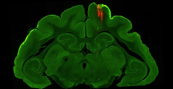
The central focus of our research is the dynamics of brain networks which we address at multiple spatial and temporal scales using neurophysiological approaches in humans and animals (ferrets). Our key goal is to relate local neural dynamics (particularly oscillations) and dynamic coupling across neural populations to sensorimotor and cognitive functions. To this end, we study the temporal dynamics of neural activity and neural interactions in the context of sensory processing, multisensory and sensorimotor integration, emotion, memory, attention and consciousness. We also aim at testing hypotheses on causal mechanisms by modulation of networks and connectivity using transcranial electrical stimulation and invasive microstimulation. Furthermore, we have a strong interest in the pathophysiological relevance of neural synchrony and change of large-scale coupling in brain disorders. In cooperation with clinical departments of the UKE, we investigate network changes in disorders such as multiple sclerosis, schizophrenia and autism.Methods and techniques
- 275-channel MEG (see Magnetoencephalography Group)
- 128-channel EEG (see Cognitive and Clinical Neurophysiology Group)15
- High density multichannel electrophysiology of single units and neuronal populations (ECoG, silicon probes)
- 7T MRI scanning (Bruker system for animal scanning, Dept. of Neuroradiology)
- Microstimulation
- Behavioral analysis
- Connectivity analysis
- Source modeling
- Histological staining methods
- Immunohistochemistry
Major collaborations
- Brigitte Röder,
University of Hamburg - Jianwei Zhang,
University of Hamburg - Christoph Herrmann,
University of Oldenburg - Peter König,
University of Osnabrück - Ilka Diester,
University of Freiburg - Thomas Stieglitz,
University of Freiburg - Pascal Fries,
Ernst-Strüngmann-Institute for Neuroscience, Frankfurt - Danica Kragic,
Kungliga Tekniska Hoegskolan, Stockholm, SE - Paul Verschure,
Universitat Pompeu Fabra, Barcelona, ES - Rainer Goebel,
University of Maastricht, NL - Karl Friston,
University College London, UK - Shangkai Gao,
Tsinghua University, Beijing, CN
-
Prof. Dr. Andreas K. Engel
-
Junior Research Group Decision Neuroscience
Head of group
- Dr. Konstantinos Tsetsos
PhD students
Research topics
Do we prefer a small flat with a short commute (A) or a large house with a long commute (B)? Many real-life decisions require combining information across different attributes. It has been shown that during such multiattribute decisions people’s preferences can be manipulated by introducing irrelevant alternatives in the choice set. For example, when a third “decoy” alternative, an even smaller flat with a short commute (C), is offered people will be lawfully biased towards the small flat with the short commute (A). These puzzling phenomena, also known as contextual preference reversals, indicate that preferences are constructed “on the fly” and that they are context-sensitive. The aim of group is to understand these phenomena at the mechanistic and neural levels. Towards this aim, we start by studying how the brain transforms sensory information into simple perceptual decisions; and building on this understanding we subsequently ask how these simple choice mechanisms scale-up in order to explain high-level choice mechanisms, such as those that dictate our consumer choices. We use a variety of methods including mathematical and computational modelling, magnetoencephalography and causal pharmacological manipulations in healthy individuals. The lab pursues the following research lines:
- Information sampling in multiattribute choice (INFOSAMPLE; funded by ERC)
- Decision irrationality and selective integration in cortical networks (research line building off a completed EU Horizon 2020 Marie Curie Fellowship)
- Temporal weighting in perceptual choices
- Risk-attitudes in decisions from experience
- Awry decision mechanisms in neuropsychiatric disease (in collaboration with Prof. Tobias Donner)
Methods and techniques- Visual and value psychophysics
- Computational modelling of cognitive processes
- MEG
- Frequency tagging
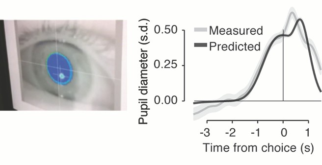
- Eye-tracking and pupilometry
- Pharmacological intervention
Major collaborations-
Tobias Donner
Department of Neurophysiology, UKE - Marius Usher
School of Psychological Science, Tel Aviv University - Nick Chater
Warwick Business School, University of Warwick
-
Cognitive and Clinical Neurophysiology
Head of group
Postdocs
- Dr. Jonas Misselhorn
- Dr. Lei Wang
- Jan-Ole Radecke
- Marius Knorr
Equipment and techniques- 128-channel EEG recording system (BrainAmp)
- Peripheral physiology recording system (skin conductance, respiration, ECG: Biopac MP100)
- Visual stimulation: 200 Hz CRT Monitor, large screen LCD projector
- Auditory stimulation: 24-channel audio surround system, EEG compatible stereo headset (E-A-RTone)
- Eyetracking (SMI iViewX Hi-Speed)
- Neuropsychological and psychopathological assessment tools
- Somatosensory Stimulation (Laser, electrical, Braille)
Autism spectrum disorders (ASD): ASD has been conceived as representing disorders of information integration or "binding" at the cognitive and neural level. In line with this hypothesis, at the perceptual level, individuals with ASD may show enhanced detail or feature perception while ignoring context or gestalt information and a superior performance for pattern parcellation or visual search. Our projects aim to investigate the relationship between potential neural and perceptual binding abnormalities in ASD.
Interaction of emotion and attention: The processing of sensory stimuli can be modulated by a variety of top-down influences. Attention and emotion are among the two most important top-down factors that continuously shape activity in sensory systems and both contribute to enhancing the saliency of sensory signals. We address the interaction of emotional and attentional processing specifically with respect to the modulation of neural synchrony in various frequency bands. We investigate how attentional and emotional factors modulate the saliency of stimuli and whether neural synchrony serves as a common underlying mechanism for these modulatory effects.
Pain processing: The broad goal of our research is to better understand the CNS pathways and mechanisms mediating pain and pain modulation in normal and pathologic states. Current research is focused on imaging changes in oscillatory activity. The dynamics and plasticity of this brain network can be studied by systematically altering somatic stimulation parameters or by observing the effects of drugs or attentional manipulations. Spatial and temporal changes in the network pattern are correlated with human psychophysical behavior to determine their functional significance.
Multisensory processing in schizophrenia: Schizophrenia with its numerous diagnostic subtypes is characterized by several behavioural deficits as well as a diverse range of cognitive, affective and perceptual symptoms, including sensory impairments. A growing body of research suggests a variety of unisensory impairments in schizophrenia at different levels of perceptual processing, from processing of stimuli of low complexity to Gestalt perception. Our research aims at investigating characteristics of neural oscillations during multisensory processing in schizophrenia.
Multisensory processing after sensory deprivation: In the developing as well as the mature brain, structural and functional changes have been observed after sensory deprivation. At the behavioural level both superior and inferior performance has been described in the remaining senses. Most studies investigating the visually deprived were concerned with unimodal processing, i.e. auditory or somatosensory processing. However, it is also of great interest whether sensory deprivation influences interactions between the senses. We investigate the role of neural synchrony for multisensory object processing and the modulation of rhythmic activity in the congenitally blind.Major collaborations
- Stefan Debener
University of Oldenburg -
Tobias Donner
University of Hamburg - Marlene Fischer
University of Hamburg - Tobias Heed
University of Bielefeld - Christoph Herrmann
University of Oldenburg - Frédéric Isel
Université René Descartes, Paris - Elizabeth Milne
University of Sheffield - Christoph Mulert
University of Hamburg - Conny Quaedflieg
University of Maastricht - Brigitte Röder
University of Hamburg - Carsten Wolters
University of Münster
-
Basal Ganglia Physiology
Head of group
Postdoctoral Fellows
Methods and techniques
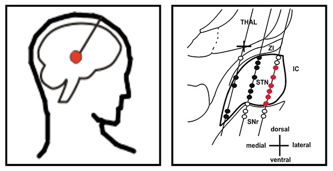
- Intraoperative microphysiology during surgery for movement disorders (therapeutic deep brain stimulation)
- Multielectrode-recordings: units and local field potentials
- Electroencephalograhy (EEG)
- Electromyography (EMG)
- Advanced time series analysis
- Experimental models of parkinsonism and tremor
- Behaviour and chronic recordings
- Experimental deep brain stimulation (DBS)
Oscillatory activity in the healthy and diseased basal ganglia: Physiological neuronal oscillations in the central nervous system cover a broad range of frequencies: from slow sleep oscillations (<2Hz) up to fast oscillatory activity in the gamma frequency range (>40Hz). These high frequent oscillations subserve a key function for physiological information processing in the healthy motor system. However, disturbances in this network, encompassing motor cortical, thalamic and basal ganglia structures, lead to pathological alterations of neuronal activity that result in the well known clinical pictures of movement disorders. We are interested in the role of such oscillatory activity in the pathogenesis of these disorders.
Cellular pathophysiology of Parkinson´s disease, essential tremor and dystonia: Pathophysiological models are derived mainly from laboratory studies. Translational research is important, since aninmal models do not fully mimic the variety of clinical phenotypes observed in human extrapyramidal disorders. However, direct access to human depth structures is limited to neurosurgical interventions. To this end, intraoperative microelectrode recordings (iMER) allow unique close-up processes of the pathophysiological alterations at a single cell level in the diseased basal ganglia. iMER increase the precision of electrode implantations for deep brain stimulation (DBS).Major collaborations
-
Wolfgang Hamel
Dept. of Neurosurgery, UKE - Edgar Kramer
Center for Molecular Neurobiology Hamburg (ZMNH) - Alexander Münchau
University of Lübeck, Dept. of Neurology - Andrew Sharott
University of Oxford, MRC Anatomical Neuropharmacology Unit - Volker Tronnier
University of Luebeck, Dept. of Neurosurgery
-
Computational Modeling and BCI
Head of group
PhD student
Methods and techniques
- 32-channel active electrode EEG systems
- systems for recording physiological signals like ECG, EMG, temperature, GSR, breathing frequency
- Robotino robots equipped with an omnidirectional drive, a webcam, and infrared distance sensors
- Lego Mindstorm robots equipped with a 2-wheel drive, an ultrasound distance sensor, a microphone, and an optic sensor
- Steady State Visual Evoked Potentials (SSVEP)
- Motor Imagery
- Attentional modulation
- Time-frequency analyses, ICA, PCA
- Classifiers
- Artificial Neural Networks (ANN), Recurrent Neural Networks (RNN)
- Dynamical systems theory
Neurofeedback: Neurofeedback allows a person to observe his or her own brain activity by giving visual or auditory feedback. This allows the user to develop strategies to adjust the own mental activity. This can be employed to bring the person into a desired cognitive or physiological state (e.g., relaxing), or to control a computer. Currently we investigate the potential of neurofeedback asa training method for BCI systems.
Computational models of oscillatory brain dynamics: Oscillatory dynamics of neuronal activity is a commonly observed phenomenon in neurophysiological experiments. The properties of networks of coupled oscillators have been investigated in a number of computational models. Current hypotheses about the functional roles of oscillations consider it as a gating mechanism, a mechanism for modulating sensory processing, or a system of internal reference clocks. Still there are many open questions about mechanisms of information processing in oscillator networks and the role of oscillations for generating behavior. Our projects in this area aim at elucidating computational mechnaisms of oscillatory networks, developing network activity analysis methods, and modelling oscillatory effects found in EEG experiments on size perception.
Computational models of cognition: As part of several cooperations supported by the EU, we are involved in projects implementing robot systems that combine visual and auditory information processing to achieve orienting behaviour, object recognition, navigation, and memory formation. The projects combine a synthetic biorobotics approach with neurophysiological experiments in humans, and computational modeling that allows to identify relevant information processing principles.Major collaborations
- Peter König
Dept. of Cognitive Science, University of Osnabrück - Shangkai Gao
Dept. of Biomedical Engineering, Tsinghua University, Beijing - Markus Werning
Dept. of Philosophy & Mercator Research Group, Ruhr University of Bochum - Björn Brembs
Dept. of Neurobiology, Free University of Berlin
-
Magnetencephalography (MEG)
Head of group
Postdocs
PhD students
Engineers and technicians
- Dr. Roger Zimmermann
- Malte Sengelmann
- Christiane Reissmann
- Gerhard Steinmetz
Methods and techniques
- 275 channel whole-head magnetoencephalography system (CTF275, VSM MedTech) with 275 axial gradiometers (superconductive quantum interference devices, SQUIDs)
- Simultaneous 64 channel EEG system (VSM MedTech)
- Online Eye tracking (SMI MEG-iView XTM)
- Visual, auditory, somatosensory and pain stimulation (Sanyo XP51 Beamer, UKM Auditory custom stimulation Querosys Piezo-stimulator, Starmedtech Thermis Laser, Ibrro RSG0405 current stimulator)
- MEG compatible button response system
- Polhemus 3D digitizer
- Time-Frequency Transformation, Beamforming, Multivariate-Autoregressive (MVAR) modelling, computational modelling, dipole reconstruction
- Analysis software: Matlab, Fieldtrip, BioSig, EEGLab, Besa, Curry
Measurement of magnetic brain signals: A super-conducting quantum interference device (SQUID) enables to detect extremely small magnetic fields originating from ongoing currents in the nervous tissue of the human brain. By means of the SQUID technology these extremely weak biomagnetic fields are measured in a non-invasive manner, even sparing the subject/patient from any physical contact with the device. In detail, MEG rests upon the physical principal that currents flowing across the dendritic membran of pyramidal neurons that constitute the cerebral cortex generate extra-cranially measurable magnetic fields. In respect to the source of brain signals, MEG is closely related to electroencephalography (EEG), which has developed to be an integral part of clinical diagnostics over the last decades. One principal advantage of using MEG is that brain activity is detected with a high temporal and spatial resolution.
Brain oscillation and synchronisation: The MEG captures neuronal oscillations generated by populations of synchronously oscillating neurons. From a functional perspective such neural oscillations might be of crucial importance, since they were shown to play a profound role for neuronal communication, exemplarily in binding object features into a coherent percept. Specifically, our work aims at investigating the idea that neuronal synchronization gives rise to the formation of representational states in human awareness and enables response selection in sensorimotor brain areas. One particular focus of our research is the visual system of the human brain, which may constitute a paradigmatic case for the mechanism of binding that integrates information processed in different, locally apart cortical areas into coherent object representations.
Tools of signals analysis such as time-frequency transformation and beam-forming algorithms allow us to locate sources of brain oscillations and their ongoing mutual synchronisation. Furthermore, by means of multivariate autoregressive (MVAR) modelling we seek to uncover the hierarchy of neuronal communication within dynamic brain networks. Various experimental paradigm and different stimulation designs including cognitive, attentional and pharmacological modulations are applied to analyse the various faces of oscillatory processes in the human brain.Major collaborations
- Laura Marzetti
University of Chieti-Pescara, Italy - Stefan Haufe
Technical University of Berlin - Forooz Shahbazi
Fraunhofer Institute HHI Berlin -
Christoph Mulert
Dept. of Psychiatry, UKE -
Claus Hilgetag
Dept. of Computational Neurscience, UKE
Downloads:
METH is a collection of MATLAB functions to conduct data analysis of MEG and EEG data. The main features are:
- EEG and MEG forward calculation in realistic volume conductors based on expansions of the lead fields in spherical harmonics.
- Inverse Calculations: Dipole fitting for ERFs/ERPs and cross-spectra, MUSIC, RAP-MUSIC, SC-MUSIC, minimum norm source reconstruction, eLoreta, and beamformers.
- Power and coupling measures: cross-spectra coherency, cross-bispectra and bicoherence, 1:1 phase locking, power correlation including corrections for artifacts of volume conduction, lagged coherence, phase lag index, multivariate coupling measures robust to artifacts of volume conduction (MIM, MIC, GIM), and wedge music.
- Decomposition: MOCA for two or more source distributions
- Statistics: false discovery rate and permutation test
- Visualization: topo-plots, head-in-head plots and time-frequency plots (all in sensor space), MRIs including results in source space, surface plots (e.g. cortex) including results on these surfaces.
This toolbox is free software without any warranty: you can redistribute it and/or modify it under the terms of the GNU General Public License as published by the Free Software Foundation, either version 3 of the License, or any later version.
