Overview
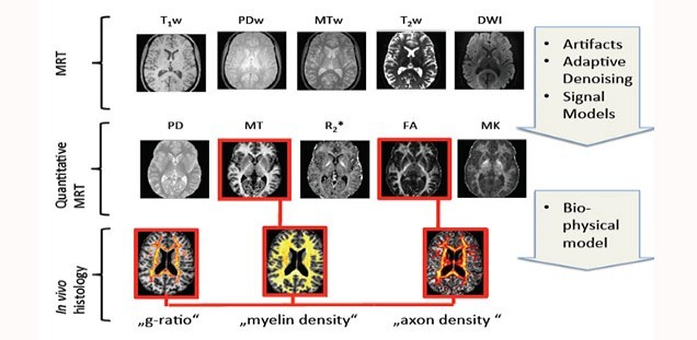
Understanding basic mechanisms underlying normal and diseased human brain developments crucially depends on reliable knowledge of its anatomical microstructure and connectivity. Our long-term goal is to estimate microstructural properties non-invasively in living subjects using biophysical models of the MRI signal.
We are developing novel methods for fusion of high-resolution quantitative MRI (qMRI) to enable MRI-based in vivo histology (hMRI) in the brain and spinal cord. The main focus is to develop valid MRI metrics that characterize key white matter microstructure properties as known from ex vivo histology (e.g. myelin density, fiber density, and the g-ratio i.e. the ratio between inner and outer fiber diameter).
To achieve these goals, we are currently working on three projects:
Basic science: Validating MRI-based in vivo histology by comparison with ex vivo histology (Funded by the DFG Emmy Noether Program )
Clinical research: Understanding the microstructural mechanisms of spinal cord injury (Funded by EU ERA-NET Neuron )
Connectomics: In vivo and ex vivo characterization of the human microstructural connectome (Funded by DFG SPP 2041 ).
Completed projects:
Marie Skłodowska-Curie Fellowship (MSCA-IF-2015): Mesoscopic characterization of human white-matter: a computational in-vivo MRI framework .
For a full publication list see:
Pubmed

