MR Physics
Overview
The MR Physics group is working on the development of new and improved methods for magnetic resonance imaging (MRI) and spectroscopy (MRS) that aim to provide a non-invasive access to the structure, organization, function, and metabolism of the human central nervous system in vivo.
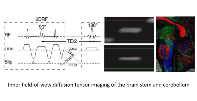
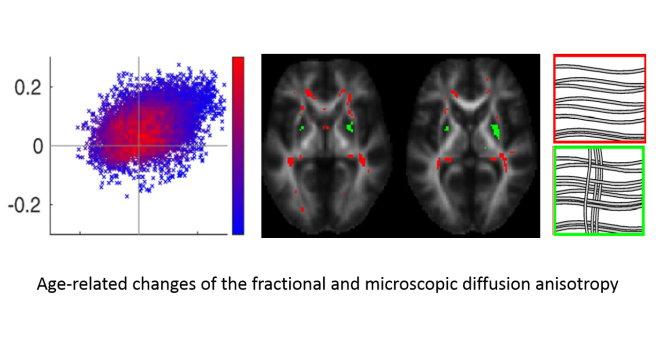
A major focus of our work is diffusion-weighted MRI, the localized measurement of water diffusion properties in tissue that are sensitive to the tissue structure on a cellular or microscopic scale. With extended diffusion-weighting methods like the double diffusion encoding used in our group, more specific information like the density, size, and shape of the cells may become accessible and could help to distinguish and characterize healthy and pathological tissue. Furthermore, we aim to speed-up and improve the image quality of diffusion-weighted MRI, in particular for applications in the spinal cord to make it clinically feasible for patients with spinal cord lesions or injury.
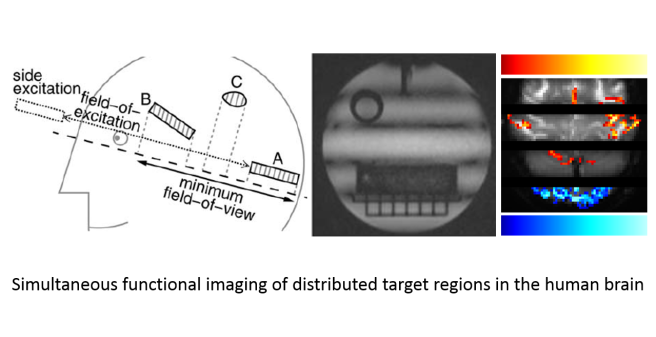
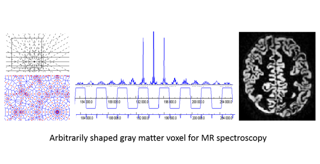
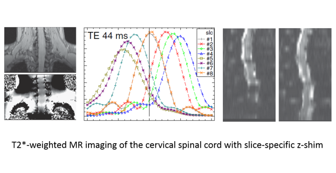
For MR spectroscopy, a technique to measure the concentrations of important metabolites, we have developed methods to acquire arbitrarily shaped target regions that can match anatomically or clinically defined target regions with minimal partial volume effects and, thus, enhance the impact and significance of the experiments. Other research projects deal with MR imaging with blood-oxygenation-level dependent contrast, the most important tool to investigate the function of the central nervous system in vivo, and aim to increase the temporal and spatial resolution and improve its applicability in the spinal cord, e.g. to investigate the processing of sensory input like pain.
In collaboration with research groups at the UKE and other universities, the developed and improved methods are applied in biomedical research and clinical studies in healthy volunteers and patients to evaluate their feasibility in practice and clinical setups. Finally, we provide methodological support for scientists performing MR experiments at the department's whole-body MR system, in particular for anatomical and functional measurements of the human brain.
